The short often slitlike passage that on both the left and right sides connects the third brain ventricle of the diencephalon with the lateral ventricles of the cerebral hemispheres.
Roof of intervertebral foramen.
The passage is bounded anteriomedially by the column of fornix and posterolaterally by the anterior pole of the thalamus.
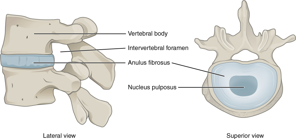
The intervertebral foramina allow passage of structures out of and into the vertebral canal.
In the intact vertebral column the vertebral foramina of all of the vertebrae align to form the vertebral spinal canal which serves as the bony protection and passageway for the spinal cord down the back.
Anterior wall of neural foramen is intervertebral disk and adjacent endplates.
In the cervical region a portion of the vertebral body below predominately the uncinate process also contributes to the anterior boundary.
A pair of spinal nerve roots leaves the dural sac just above the level of each intervertebral foramen.
Roof inferior vertebral notch of the vertebra above.
Posterior wall is facet joint and inferior and superior articular processes.
The intervertebral foramen also called the neural foramen is the opening between the vertebrae through which spinal nerve roots travel and exit to other parts of the body.
This sleeve encloses the nerve roots as far as the intervertebral foramen and spinal nerve where the dura.
A number of structures pass through the foramen.
The large opening between the vertebral arch and body is the vertebral foramen which contains the spinal cord.
Roof and floor of neural foramen are formed by pedicles of adjacent vertebrae.
The intervertebral foramina which transmit the spinal nerves and the accompanying radicular arteries which supply the spinal cord are on the lateral aspect of the vertebral column.
The word foramen is the singular form while foramina is the plural form.
Anterior wall cervical and lumbar regions are the same however because of the size and the fact that the ribs are in the thoracic the thoracic wall will differ.
They do so by penetrating the dural sac in an inferolateral direction taking with them an extension of dura mater and arachnoid mater referred to as the dural sleeve see fig.
The foramina or openings are present between every pair of vertebrae in these areas.
Each foramen lying between the pedicles of the adjoining vertebrae is bounded anteriorly by the vertebral bodies and the disc and posteriorly by the facet joints fig.
In the lumbar region where nerve root compression is more common the nerves increase in size from above downwards whereas the.
Anterior lower posterolateral aspect of a vertebral body and the intervertebral disc below in the thoracic and lumbar regions.
Nerve roots dorsal root ganglia and dura.
Also because the.
Floor superior vertebral notch of the vertebra below.
If the foramina narrow they can put pressure on the nerve roots near them causing pain.
Cervical thoracic and lumbar vertebrae all have intervertebral foramina.